Library
Frequently Asked Questions
Some horses will require ongoing management, while others may experience various periods of improvement. PetDerm will work closely with your primary veterinarian to create a responsive and malluable treatment plan to match your horse’s current symptoms.
Autoimmune skin conditions are usually managed rather than cured. With the right treatment plan, many horses can remain comfortable, and active. Early detection can help slow the progression of the disease.
They can be uncomfortable or painful, especially when lesions are inflamed or slow to heal. Proper management focuses on reducing discomfort and preventing complications. PetDerm’s dermatological veterinarian provides an individualized management plan to improve the overall quality of life for your horse.
Yes! Reducing exposure to dust, mold, and airborne irritants in barns and stables is one of the most effective ways to manage equine asthma. Especially in Alberta’s harsh climate where horses spend more time indoors during the winter months.
Equine asthma is often managed long-term rather than a curable disease. With appropriate environmental control and allergy management, many horses can have a comfortable and improved quality of life. PetDerm’s goal is the provide you and your primary veterinarian with a sustainable long-term management plan.
No. Equine asthma is not caused by bacteria or viruses. It is an inflammatory condition often linked to allergies and environmental exposure, which is why antibiotics are not typically effective. A dermatologic assessment may be necessary to evaluate the underlying cause and early on intervention to slow the progression of the disease.
Early signs may include occasional coughing, reduced stamina, slower recovery after exercise, or subtle breathing changes. These signs can be easy to miss at first but may worsen over time without medical intervention. If you suspect that your horse has enviornmental allergies, ask your primary veterinarian for a referral today!
In many horses, equine asthma is part of a broader allergic response. The same allergens that cause skin problems can also affect the airways, which is why some horses experience both skin and respiratory symptoms.
Learn more about PetDerm’s intradermal allergy testing.
Yes. Adjustments such as improving regular hygiene, reducing moisture exposure, managing insects, or modifying stable conditions can play an important role in preventing flare-ups and supporting treatment success.
Some equine skin conditions can be ongoing and require long-term management rather than a one-time treatment. With proper care, many horses can remain comfortable and active even with chronic skin disease.
They can. Skin pain, irritation, or lesions—especially in areas under tack or around the legs—can cause discomfort, behavioral changes, and reduced willingness to work. Managing skin disease helps support both comfort and performance.
You should consider a dermatology veterinary evaluation if a skin problem:
- Persists longer than expected
- Keeps returning after treatment
- Causes significant itching or discomfort
- Spreads or worsens over time
- Interferes with riding, training, or daily comfort.
Early assessment can help prevent minor issues from becoming chronic.
Horses are constantly exposed to moisture, insects, dirt, allergens, and changing weather conditions. These factors can contribute to weakening the skin’s natural barrier and making it easier for irritation, infection, or allergic reactions to develop. Most frequently seen on the body like the legs, mane, tail, and face.
It is case dependent on your horse. In some cases, pasture access may need to be modified or changed, as grasses can act as dietary triggers for certain horses. This will depend on your horse’s history and symptoms. PetDerm’s team will review in your consultation what is resonable and accessible for you and your horse, if an elimination diet trial is suggested.
Currently, there is no single blood or skin test that can reliably diagnose food allergies in horses. The most accurate method is a carefully managed elimination diet followed by controlled reintroduction of foods to identify the trigger. The PetDerm can provide guidance on an elimination trial while providing immediate support to your horse’s gut health.
Yes. A horse can develop a reaction to a food they have previously tolerated well. Over time, the immune system may begin reacting differently to certain ingredients, leading to new or worsening symptoms.
Immunotherapy works by gradually desensitizing the immune system to identified allergens, helping reduce the severity of allergic symptoms over time.
-
Long-term control of allergic skin disease
-
Reduced reliance on symptomatic medications
-
Improved comfort and quality of life
-
Management tailored to Alberta’s environmental allergens
Immunotherapy is particularly beneficial for horses requiring ongoing allergy management and those with limited response to standard treatments.
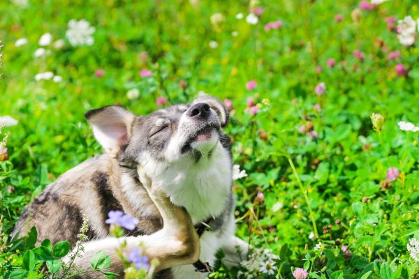
Horses with environmental allergies may show a wide range of symptoms, including:
-
Persistent or seasonal itching
-
Hair loss or broken hair coat
-
Skin thickening or pigment changes
-
Recurrent hives (urticaria)
-
Crusting, scabbing, or oozing lesions
-
Secondary bacterial or fungal infections
Because these signs can overlap with other skin conditions, accurate diagnosis is essential before initiating long-term therapy strategy.
Calgary’s PetDerm internal medicine vet is a board-certified Veterinary Internist. They complete an additional 4 years of dermatology or internal medicine residency before they become board-certified. The residency program involves extensive training in anatomy, physiology, pathology, clinical diagnosis, research, and more. A board-certified veterinarian dedicates at least 10 years to education and residency, similar to a doctor in human medicine.
A pet Internal Medicine specialist is trained in diagnosing and treating a broad spectrum of internal health issues in animals. They are highly trained and experienced in managing gastroenterology, endocrinology, infectious disease, urology, nephrology, respiratory medicine and hematology & immunology. It is common for a patient’s disease to impact multiple organs, requiring a detailed understanding on how to balance multiple issues for the best quality of life.
Whether it’s endocrine disease, gastrointestinal troubles, autoimmune problems, or chronic organ disease, our team works closely with your primary-care veterinarian to provide thorough diagnostics, personalized treatment plans, and ongoing support.
It’s easy to assume snoring or noisy breathing is “normal” for short-nosed dogs, and BOAS is often missed until it becomes advanced. The RFGS provides:
- An objective way to classify breathing ability
- Early detection of airway obstruction
- A clear guide for whether surgery would help
- A baseline to track improvement after treatment
Clinics using RFGS can confidently determine whether a dog would benefit from procedures like laser BOAS surgery, and how urgently.
The RFGS is a standardized way to measure how well a brachycephalic dog can breathe. It was developed by experts at the University of Cambridge to provide a consistent, objective way to diagnose BOAS and determine how severe it is.
It helps identify dogs who need treatment before they reach a crisis stage.
The RFGS includes two main parts:
-
Listening to breathing at rest
The veterinarian listens for signs like snoring, wheezing, stridor (high-pitched breathing), or excessive panting. -
A short, controlled exercise test
Your dog walks or trots on a lead for a few minutes (usually around 3 minutes).
After the walk, the veterinarian evaluates your dog’s breathing again — checking recovery time, airway noise, and effort.
This gentle test is safe and designed to mimic normal activity your dog would experience at home. A pre-screen chest x-ray may be required.
After surgery, rest and limited activity are essential. Monitor your pet’s breathing, attend follow-up visits, and follow all post-operative care instructions. Dogs often experience less pain and recover faster after laser surgery. Post-surgery Phovia Treatment may be offered to further expedite safe, bacteria-free healing.
Only Grade 0 dogs are considered BOAS-negative. Grades 1–3 are BOAS-positive with increasing severity.
In North America, there are common breeds that have a higher incidence of BOAS due to exterme brachycephalic anatomy:
- French Bulldog
- English Bulldog
- Pug
- Boston Terrier
- Pekingese
- Shih Tzu
These breeds can experience BOAS if their individual conformation is more extreme (i.e short muzzle, narrower nostrils, and elongated soft palate):
- Boxer
- Cavalier King Charles Spaniel
- Lhasa Apso
- Brussels Griffon
- Japanese Chin
Your family veterinarian may start by gathering a detailed history about your dog’s breathing habits – snoring, snorting, tiring quickly, heat intolerance, regurgitation, or noisy breathing. They will perform a full physical exam.
If BOAS is suspected, ask your family veterinarian to refer your pet to PetDerm. Dr. Becky Valentine does a detailed assessment using the Respiratory Function Grading Scheme (RFGS).
Even after successful surgery, long-term care is vital. Maintain a healthy weight, avoid overheating, encourage hydration, and engage in gentle exercise. These steps help prevent recurrence and support lasting respiratory health.
Over time, BOAS can lead to other health issues as a dog is working much harder to move air. Some conditions may appear alongside BOAS:
- Weakening of the voice box (laryngeal collapse) – When a dog struggles to breathe for a long time, the cartilage in the throat can weaken and start to collapse inward, making breathing even harder.
- Lower airway collapse (bronchial collapse) – If the upper airway becomes very compromised, the smaller airways deeper in the chest can also start to narrow or collapse.
- Digestive issues – Dogs with BOAS often swallow more air when trying to breathe, which can lead to problems like reflux, vomiting, or even a sliding stomach (hiatal hernia). Many of these digestive issues often improve once their airway is corrected.
- Aspiration pneumonia – Because breathing and swallowing become more difficult, there is an increased risk of food or liquid accidentally entering the lungs, which can cause a serious infection.
Common symptoms include loud snoring or breathing, exercise intolerance, gagging or coughing, cyanosis (bluish tongue or lips), and heat intolerance. Recognizing these signs early is crucial as untreated BOAS can progress to severe respiratory distress in up to 40% of cases.
Endoscopy is a minimally invasive procedure where a tiny camera is inserted into your pet’s digestive or respiratory tract to see inside and take tissue samples (biopsies) if needed.
Endoscopy helps veterinary internists diagnose causes of vomiting, diarrhea, weight loss, coughing, or difficulty swallowing. It’s often used to identify inflammation, infections, or foreign objects.
Yes. Endoscopy is generally very safe. It avoids the need for major invasive surgery and usually involves light sedation.
Endoscopy is a less invasive procedure, has a faster recovery time, causes less discomfort, and reduces the risk of complications compared to open surgery.
Most pets can go home the same day, though the PetDerm team may recommend a short observation period depending on their health and the procedure performed.
Visual findings are often available immediately. If biopsies are taken, results typically come back in a few days. PetDerm’s veterinary internist will discuss the samples and testing timelines with you prior to the procedure.
Yes. Veterinary X-rays use very low radiation levels, and exposure is kept to the minimum necessary to get a clear image.
Usually, no special preparation is needed. The PetDerm team will let you know if fasting is required if sedation is planned for. A team member will discuss this with you when booking the diagnostic procedure.
Most X-rays are quick—often just a few minutes. It may take longer if multiple views are needed or if your pet requires sedation.
Images will be reviewed by a veterinary radiologist for a more detailed report and PetDerm’s veterinary internist will follow up with you.
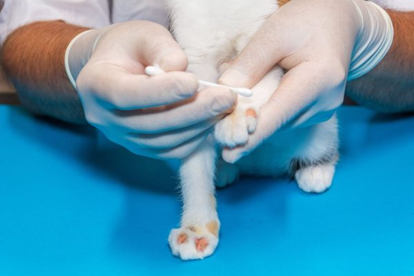
Yes. When indicated, we can perform ultrasound-guided fine needle aspirates (FNA) or biopsies to obtain tissue or cell samples for further diagnostic testing.
Cytology involves collecting cells using a fine needle and examining them under a microscope. This is minimally invasive and provides rapid preliminary information about the nature of a mass or organ change—whether it is inflammatory, infectious, or potentially neoplastic.
A fine needle aspirate (FNA) is performed using a thin needle, often without the need for general anesthesia, to collect a small number of cells from an organ or lesion. This is useful for screening and staging, particularly in cases involving the liver, spleen, lymph nodes, or abdominal masses.
Preliminary findings are often discussed the same day, with a detailed interpretation provided shortly thereafter by our board-certified veterinary radiologist.
Most ultrasounds take 30–60 minutes, depending on the complexity of the case and whether additional procedures (such as fine needle aspirates) are needed.
Yes. The area of interest is shaved to allow the probe to make direct contact with the skin and achieve the clearest images.
Fasting for 8–12 hours is typically recommended to reduce interference from gas in the intestines. Water is generally allowed. Specific preparation instructions will be provided by a PetDerm technician when your appointment is scheduled.
Yes. Ultrasound does not use radiation and is considered safe, lower risk procedure. Most pets do require sedation to allow for precise imaging position.
Ultrasound is a non-invasive imaging technique that uses sound waves to create real-time pictures of your pet’s internal organs. It helps assess their structure and detect abnormalities without surgery.
It is not recommended to use raw food for an elimination diet trial. Raw diets can pose health risks due to potential contamination with harmful bacteria like Salmonella and Listeria. These bacteria can make both pets and their owners’ sick. For a safe and effective elimination diet, it’s best to use a specially formulated commercial diet or a home-cooked diet prepared under the guidance of a veterinarian.
Food allergy is a genuine allergic response where the immune system becomes overly sensitive to ingredients a pet has previously eaten. For food allergy to occur, the pet must have been exposed to the problematic allergen or a similar one with cross-reactivity. On the other hand, food intolerance doesn’t need prior sensitization and can happen on the first exposure. In dogs and cats, food intolerance reactions typically manifest as gastrointestinal issues like diarrhea or vomiting.
An elimination diet for a cat or dog involves feeding them a special diet that excludes all the ingredients they have previously eaten. This helps identify which foods might be causing allergies. The process typically lasts for at least 8 weeks, but it can extend up to 12 weeks. During this time, it’s crucial to avoid giving your pet any treats, chews, or flavored medications that could interfere with the trial. If symptoms improve, potential allergens are reintroduced one at a time to pinpoint the specific cause of the allergy. The PetDerm team can help you develop an elimination diet to improve your pet’s quality of life.
The only reliable way to diagnose food allergies in dogs and cats is through a strict elimination diet trial. While some tests claim to detect food allergies using blood, hair, or saliva, they are not reliable or dependable.
Symptoms of food allergies in dogs and cats may include behaviours such as licking, scratching, chewing, and rubbing various regions like the face, ears, rear, armpits, and other body parts. Additionally some pets with food allergies may present gastrointestinal manifestations such as loose stool and vomiting. Distinguishing food allergies from environmental allergies can be complicated and that is why we are here to help. Diet consultations with a veterinary dermatologist can help your pet enjoy food again!
Food allergies account for up to approximately 30% of pets with allergies, but they can be layered with allergies to house dust mites, pollens, grasses, and trees, which makes it hard to treat with diet alone. And because there is no accurate food allergy testing available in veterinary medicine, a change in diet might only work when addressing other factors that could be causing your pet’s rash, itch, or ear infection.